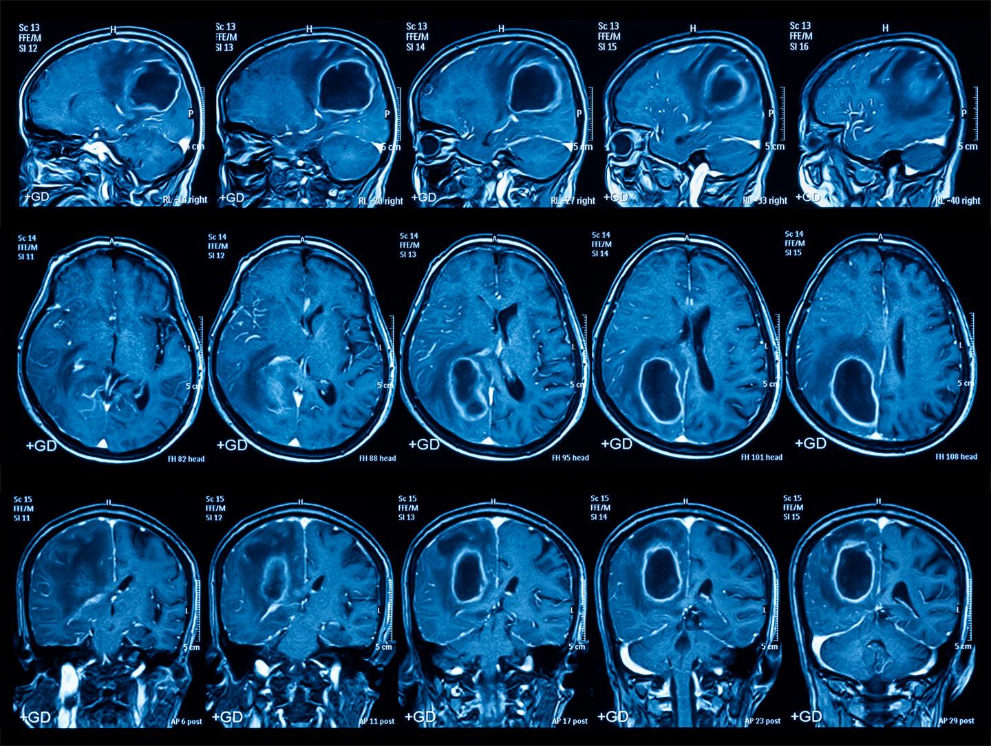
CERN’s Know-How
- Design and characterization of detectors for applications that require fast imaging and high spatial resolution. Applied to nuclear medicine imaging and radiology.
- Hybrid pixel detector technology was developed for particle tracking at CERN and has enabled high resolution, color X-ray CT imaging.
- Significant know-how in the design, integration and testing of state- of-the-art fast, low noise, and radiation-hard multichannel electronics.
- In-house design and modelling of superconductive devices and magnet systems and ‘one-stop-shop’ with a range of unique facilities for testing ideas and innovative concepts.
- Software frameworks and expertise for calculations of particle transport and interactions with matter, used to simulate imaging detectors. Significant experience in data storage, data analysis and image reconstruction software.
Facts & Figures
- 1977: First PET image taken at CERN.
- 2018: First 3D colour X-ray image of the living human body taken by MARS Bioimaging, using Medipix technology.
- 20 years of development on Medipix.
- 7 tools for medical applications have been based on GEANT4 simulation toolkit.
Value Proposition
Read more about Medical Imaging here.

